


Onychomycosis is a type of fungal lesion, which exclusively affects the surface of the nails.In order to avoid the propagation of infection over time, you should know what the fungus of the nails looks like, because the therapeutic effect will be reached more quickly if the treatment started at an early stage of the infection.Most often, the disease occurs in the elderly due to low immunity, but the risk of obtaining a disease concerns each person.There is no unique classification of fungal nails damage;In medical practice, it is usual to distinguish them instead of the location and the depth to which they penetrate.The infection is also grouped according to the type of pathogen.
Types of pathogens and primary signs
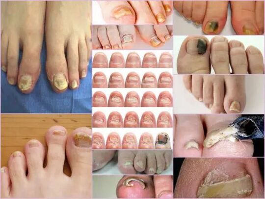
Symptoms of the nail fungus at the initial stage are easily determined directly by nail condition.This sign is the most informative, because onychomycosis is always manifested in the form of changes in the color of the surface, the deformation of the bed, exfoliation and any external change.The latter are expressed by roughness, the formation of grooves and cracks, as well as by interspersed and general violation of the nail.
The main sign of a healthy nail is pink and transparency.Onychomycosis at any stage is characterized by the turbidity of the nail and the color change in yellow, brownish or greenish black.At an advanced step, the surface can acquire a black shade in the background of almost complete destruction.
The external signs of infection by a fungus depend on the type of pathogen that has infected.In medical practice, the following possible lesions are distinguished:
- Candida infection by a fungus, which is expressed by the discharge of the nail directly at the base of the box.Candidiasis of the rollers around the nail will be characteristic of candidiasis.This version of the complication can have a bacterial source in the form of streptococci or staphylococci, either expressed in the form of medium plates or psoriasis;
- Dermatophytes of the Trichophyton Rubrum type.In this case, the penetration of the infection occurs directly from the free edge of the nail.The first symptoms of such a pathogen are the appearance of a yellowish stain on the surface.In the field of neoplasm, the nail collapses and the spot itself tends to increase.The common location of the neoplasm is along the plate, parallel to nail rolls, in this case, the infection is called lateral distal.There is another form of defeat by this pathogen - distal, in which the stain appears on the part removed from the hole, mainly in the middle of the free edge;
Important!
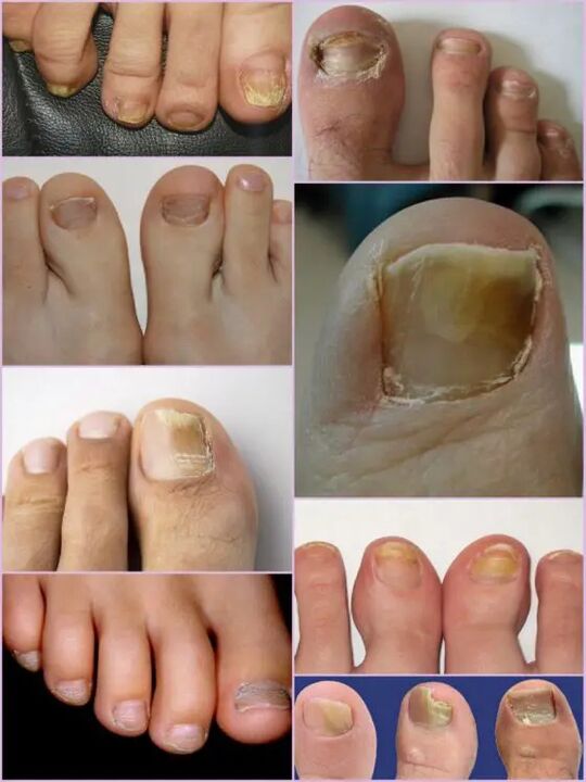
This type of fungus occurs most often on the thumbs of the legs, gradually transforming the nails into a loose yellowish mass.In the absence of appropriate treatment, the disease turns into hyperkeratosis.The nail plate is completely destroyed due to the spread of the infection around the perimeter.
- Dermatophytes like Trichophyton Mentagrophytes.Onychomycosis with such excitators is also most often based on thumbnails, less often than little fingers.An infection with a fungus of this type requires therapy not only of the nail, but also feet due to the rapid spread of the pathogen.A symptomatic disease can be like Leikonichia, a common disease in medical practice.The main signs are white spots that appear on various parts of the nail, neoplasms are distinguished by a non -standard form and different sizes.It is easy to distinguish the fungus from the Lakonichie - in the latter case, spots are an accumulation of air, which is not observed with fungal damage;
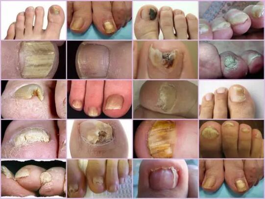
- Fungus mold.This damage option is much less often than a candida or dermatophytic form.The main sign of such an infection - the surface of the plate acquires a dark, almost black shade.Not the whole finger can be fully infected, but only one part of the nail plate.The first signs of nail fungus on the legs of this type are a clear change in color.Onychomycosis can develop in the form of a longitudinal strip of black or dark green on the bottom of the rest of the pink part of the nail.
Diagnostics by type of change
It is not difficult to notice a fungus of the nails on the legs even at the primary stages of the infection, because an infection of this type manifests itself quite actively from the first day of the lesion.Instead of the usual transparent nails of pale pink in the patient, a significant surface deformation and a general change in the disease are observed.The affected area has a dull yellowish shade, which appears mainly on the thumbs.The type of fungus and the degree of damage are determining factors.
At the first step, fungal damage to the legs on the legs are the appearance of small households.The thickening of the plate and the keratinization of the bed under the nail will be characteristic.This step is accompanied by such a phenomenon as partial detachment and discharge of the nail plate, which serves as a source of infection for healthy people.
Despite the active thickening of the plate, its constant grinding can be observed, whatever the current factors.The characteristics of each step and symptoms of the fungus on the legs depend directly on the type of pathogen.
According to the modifications that occur with the nail plate, three options for onychomycosis are distinguished:
- Hypertrophic;
- Normotrophic;
- Atrophic.
In the first case, there is a clear change in the shadow of the nail plate, its destruction along the edges, as well as the deformation of the surface of the plate.The nail thickens so much that it causes feelings of discomfort and painful during walking.Normotrophic nail nail mycosis is distinguished by the presence of a healthy shine, but the plate itself acquires a little yellow, spots form on it.With an atrophic type of damage, brown and gray households are formed on the surface of the nails.Thanks to them, it is possible to precisely determine the location of the pathogen.
Important!
The type of atrophic or onycholithic nail fungus is characterized by thinning of the nail plate, not its growth, as in other cases.The areas on which the pathogen is located tend to detach.The lack of appropriate treatment leads to an advanced stage - the complete rejection of the nail plate.
Location classification
Another sign by which you can separate the fungal damage to the toes from the toes depends directly on the location of the households on the nail plate.This also includes the depth of the pathogen, which in turn allows you to determine the approximate duration of future treatment and the chances of rapid recovery.
Fungal diseases of leg nails on the location of the location are classified in the following groups:
- Onychomycosis is a type of white surface - the appearance of many whitish spots on the surface of the nail plate.A fungal infection leads to detachment of the skin to the places of appearance of the spots on which there is an active discharge of ladders.The advanced stadium leads to the destruction and complete rejection of the plate;
- Distal - develops on the free edge of the nail.The color change is first observed on a small area of the plate, after which there is an active expansion of its limits.The lesion is characterized by a yellowish or brown gray shade, as well as an unequal rough surface and progressive exfoliation;
- Lateral onychomycosis has the development stages similar to distal, but it is located exclusively on the side sides of the nail plate;
- Total infection - complete infection of the entire surface;
- Proximal onychomycosis.This disease begins its activity from the cuticle, which is inflamed, then the fungus quickly affects the nail, and the process itself begins with the appearance of a small white spot near the inflamed area of the periolian roller.
The most common forms of nail mycosis on the legs are lateral and distal, which are generally caused by dermatophytes.Such forms of damage such as proximal and white can act as a secondary disease that accompanies the disease of the immune system, for example, VICH.Total nail damage with a fungus must be considered as an advanced stage of development of any fungus under the nail.
Characteristics of the fungus under the microscope
Despite the presence of an impressive number of signs of onychomycosis, other ailments associated with skin problems and not infected in nature are accepted for fungal damage to legs.You can only determine the exact diagnosis and the type of pathogen only in the laboratory under a microscope, for which, in a hospital, a scraping of biological equipment in the affected areas is carried out.
The resulting biomaterial is pre-plated in an alkaline environment, after which a multiple increase is made.The study of the fungus of the nails under a microscope allows you to see an active pathogen, whose external shape determines its type, the distribution scale and a degree of approximate damage.On a nutritious environment, you can predict the approximate growth of the colonies, which not only makes it possible to make a precise diagnosis, but also to determine the limitation of the infection.
Important!
Since it is possible to determine the presence of onychomycosis only in a laboratory, you should not postpone a visit to the doctor at the slightest suspicion.The nail fungus develops quickly and the waste of time increases therapy.
What is an alarming signal?
The nails mushroom on the legs are manifested by certain symptoms which are partially similar to certain skin conditions.The following signs are more likely to indicate an onychomycotic lesion which requires medical intervention:
- The appearance of yellow spots on the plate, its deformation, a change of structure, which was not observed earlier;
- The darkening of the plate, the loss of transparency, certain photos of the fungus of the nails on the legs show a shade near the black, which is characteristic of the mussels of the pathogens;
- Thickening and keratinization of the nail plate, or vice versa too clear of slimming;
- Rejection of the bed nail, detachment of scales and crumbs;
- Swelling of the roller, the suspension above the plate, release of liquid;
- The nails affected by the fungus look inflamed, regardless of the type of pathogen.
All of these symptoms indicate a high risk of infection.Certain external symptoms of modifying the structure of the nail plate may be the result of other diseases.With increased fragility, the quantity of calcium and iron in the body must be increased, an increase in tuberosity means that any body infection, with scratches and purely white points, a lack of copper or zinc is possible.Despite the fact that in the photo, the fungus of the nails looks like focal damage on the plate with a color change closer to yellow, this does not always indicate the infection.Yellow can indicate various problems with the stomach and the liver.
It is not difficult to recognize onychomycosis on the fingers by external signs, despite the in -depth classification of the disease.But you can determine the exact type of the pathogen and the stage of damage can only be in the laboratory.